The optical analysis of structures in mammalian cells, in particular in human cancer cells, is essential for a mechanistic understanding of the cell answer to therapeutic treatments, such as irradiation or the use of chemotherapeutic agents. For example, cellular effects in DNA damage repair and cell communication, which occur on the nanometer scale, can only be studied using high-resolution optical analysis.
Due to the diffraction limit of light, conventional optical epifluorescence microscopy with a maximum resolution of about 250 nm is only suitable for these analyses to a limited extent. This fundamental limit has already been described by Ernst Abbe as early as the 19th century. However, with the development of modern microscopy methods, scientists have found ways to circumvent this limitation and achieve resolutions from 100 nm to a few 10 nm. One of these methods is STED (stimulated emission depletion) microscopy. Here, the spatial superposition of excitation laser and doughnut-shaped de-excitation laser is exploited to improve the resolution to below 100 nm. Stefan W. Hell was awarded the Nobel Prize for this invention in 2014.
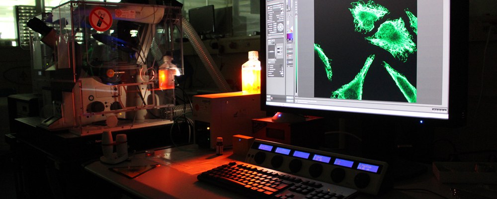
The commercial Leica TCS SP8 3X STED (stimulated emission depletion) microscope installed at the LRT2 allows us to follow the accumulation of repair proteins during DNA double-strand break repair in detail on the nanometer scale and thus to study the interactions between various proteins and with the DNA.
The microscope also has an installed climate chamber with CO2 supply, which enables studies on living cells. The high-resolution live-cell microscopy enables insights into network formation among different cells and cell-cell communication via nanometer-sized cell membrane channels and thus to witness the interconnectedness of cells. Furthermore, it is possible to follow and analyze structural changes of cell organelles live.
We offer the use of our STED microscope for studies of both fixed and living cells for potential users in the field of radiation and cell biology. A bio-lab with all necessary equipment for microscopic experiments, cell cultures and staining and microscopy protocols can be shared. The user support staff are experienced in developing new protocols as required, and in implementing and performing these protocols in collaboration with the external users.