At the Munich particle accelerator, the unique and most sensitive 3D hydrogen microscopy of thin films (30 µm (tungsten) - 180 µm (silicon)) is installed, using proton-proton scattering. Here, a proton beam with an energy of up to 30 megaelectronvolts is focused on the sample. The protons scatter from the atoms of all elements as they pass through the sample. However, when a proton strikes a hydrogen nucleus, i.e., also a proton, a special scattering reaction occurs. Comparable to the balls of equal mass in a classical billiard game, the two particles scatter apart with the precisely defined angle of 90° because of the equality of mass. The scattering event with this angular correlation can be detected with the help of a structured detector in a temporal coincidence and the background can be almost completely eliminated by other scattering events. The scattering probability for the proton-proton scattering process is known very exactly and with a known number of the injected protons it is possible to determine the hydrogen content absolutely without material-dependent calibration of the measurement.
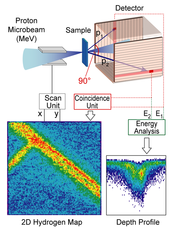
At the scanning ion microscope SNAKE a lateral resolution of ~1 µm is achieved while the depth resolution of the method is in the range of a few micrometers. On polycrystalline diamond, even a detection limit of less than 0.08 at-ppm (8 hydrogen atoms per 100 million carbon atoms) has been achieved so far [P. Reichart et al., Three-Dimensional Hydrogen Microscopy in Diamond. Science, 306 (2004) 1537]. (Download: FREE pdf, supporting online material). For further publications see the list of publications of the institute.